
Probing the Neural Basis of Visual Working Memory in Early Development
This study examined the early development of visual working memory. To do this, we studied two cohorts of children. Cohort 1 was enrolled at 6 months of age and returned for testing at 18 months and 30 months of age. Cohort 2 was enrolled at 30 months of age and returned for testing at 42 months and 54 months.
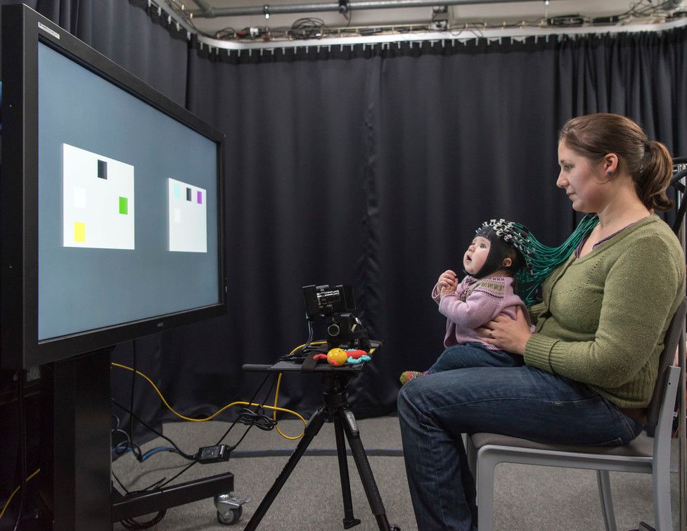
At each data collection time point, families came into the lab at UEA for two sessions. During these lab sessions, children completed a preferential looking visual working memory task while we measured brain activity using optical imaging (functional near-infrared spectroscopy -- fNIRS). At 30 months of age, children completed the Minnesota Executive Function Scale. At 42 and 54 months, children completed a change detection task -- an assessment of visual working memory commonly used with older children and adults. We also asked parents to complete several questionnaires, and we recorded parents and children playing together to look at the quality of social interactions.
Children also completed a home visit each year where we recorded parents and children playing together. In addition, families used two devices in the home. One recorded sleep behaviour over several days. The other was an audio recorder that tracked the amount of language input to children over several days. Finally, infants completed a structural MRI scan at the local hospital.
In addition to these sessions, all children completed a follow-up appointment at home at 78 months of age. We measured language skills, maths skills, executive function abilities, and attentional abilities. Parents completed several behavioural questionnaires. We also obtained SATS scores from schools as available.
Our central goal was to describe how the visual working memory system develops and how this early-developing cognitive system is related to the emergence of other abilities including later executive function and language skills. We made several key discoveries.
First, our data show that individual differences in visual working memory (VWM) abilities are stable longitudinally, that is, VWM abilities at time 1 predict VWM abilities at time 2. Furthermore, VWM abilities at time 2 predict VWM abilities at time 3. This is important, as it suggests the VWM task can be used as an early assessment tool to predict later cognition. There are not many tasks that can be used consistently across the age range from 6 to 54 months, so this finding could be important for early assessment.
Second, we identified a brain network of 7 regions that underlie performance in the visual working memory task during early development. These 7 regions showed changes in patterns of activation and suppression as the memory load varied in the task. Importantly, some brain regions showed different patterns of activity for children with less educated mothers. Maternal education is often used as an index of the socio-economic status of a family. Children with a less educated mother showed patterns of brain activity consistent with heightened distractibility and less mature brain responses as the working memory load was varied.
Another critical finding from our study is that early VWM measures predict later executive function skills. Executive function is a key skill that develops rapidly between 3 and 5 years and helps children regulate their own behaviours. Importantly, executive function skills in this early period are predictive of longer-term educational outcomes. A key question is whether we can predict which children will have strong executive function skills and which children are at risk for executive function challenges. Our data show that VWM abilities at 6 and 18 months predict executive function skills at 30 and 78 months. This might help us identify ‘at risk’ children in infancy which could guide early intervention efforts.
Our data set is unique in that we obtained longitudinal measures of how brain myelin changes in early development. We used these data to track how the structure of the brain changes in the context of pervasive experiences including language exposure. Our data show that early exposure to a large amount of adult language input delays the formation of myelin in language processing areas of the brain. Importantly, by 30 months of age, this pattern of delayed maturation creates a rebound effect such that children exposed to more language input have more myelin than children exposed to less language input. To our knowledge, this is the first demonstration that enriched input in infancy can lead to initially delayed maturation followed by a greater myelin baseline after the second year of life.
Our research was funded by:
Publications:
Johns, E., Forbes, S., Delgado Reyes, L.M., Buck, C. & Spencer, J.P. (2026). Tracking the trajectory of executive function from 2.5 to 6.5 years of age and the impact of COVID-19. Child Development, 1-17, https://doi.org/10.1093/chidev/aacag002.
Aneja, P., Kinna, T., Newman, J., Sami, S., Cassidy, J., McCarthy, J., Tiwari, M., Kumar, A. & Spencer, J.P. (2024). Leveraging technological advances to assess dyadic visual cognition during infancy in high- and low-resource settings. Frontiers in Psychology, 15:1376552. doi: 10.3389/fpsyg.2024.1376552.
Fibla, L., Forbes, S.H., McCarthy, J., Mee, K., Magnotta, V., Deoni, S., Cameron, D. & Spencer, J.P. (2023). Language exposure and brain myelination in early development. Journal of Neuroscience, 43(23), 4279-4290, doi.org/10.1523/JNEUROSCI.1034-22.2023.
Forbes, S.H., Wijeakumar, S., Eggebrecht, A.T., Magnotta, V.A. & Spencer, J.P. (2021). Processing pipeline for image reconstructed fNIRS analysis using both MRI templates and individual anatomy. Neurophotonics, 8(2), 025010, doi: 10.1117/1.NPh.8.2.025010.
Spencer, J. P. (2020). The development of working memory. Current Directions in Psychological Science, doi/10.1177/0963721420959835.